Ultrasound During Pregnancy: What Are The Risks and Benefits?

Heather Dessinger

This post contains affiliate links.
Click here to read my affiliate policy.
Ultrasound During Pregnancy: What are the risks and benefits?

Two pink lines. I remember where I was standing the first time I saw them, and how I immediately started looking forward to my first ultrasound. I wanted to know everything was okay and connect with this little being that was going to grow to the size of a watermelon and . . . well, let’s just say that as my train of thought continued along those lines I completely forgot about the ultrasound for awhile.

When I did finally mention it to a friend, she gently suggested that I might want to research a little more before getting one without a clear medical need to. I did, and what I learned surprised me. Here’s why:

  • Routine prenatal ultrasounds have not been shown to improve outcomes for mothers or babies. (1) (2) (3) (4)
  • No major epidemiological studies have been conducted on the safety of prenatal ultrasound within the U.S. in over 25 years.

Before we go any further, know this: I am not against the use of prenatal ultrasounds and think there are times when they are incredibly valuable. With that said, every mama should be fully informed about the potential risks and benefits of having one, or two, or six based on her unique situation. That’s why in this post we’ll cover:

  • What peer-reviewed research says about the safety of ultrasound
  • The effect of routine ultrasounds on birth outcomes
  • What conditions ultrasounds can screen for (and alternative test options when available)
  • Steps to limit exposure when ultrasounds are needed or wanted

After reviewing the research I chose to be selective about the use of ultrasound during my pregnancies, opting for one only if medically indicated. Your approach may be different, and I completely support that.

Also, please keep in mind that “Boo Boo Kisser” is about as official as things get for me professionally. I am not a doctor and this is not medical advice. Now let’s dive in.

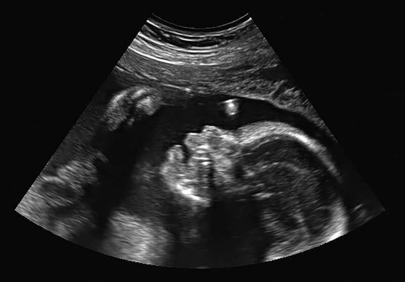
What is a prenatal ultrasound? ^

Originally developed by the military to detect submarines via SONAR, ultrasound technology bounces ultra high frequency sound waves off the soft tissues and bones of a baby in the womb, then uses the pattern created by the different surfaces to generate an image.

What is a prenatal ultrasound?

What’s the difference between a sonogram and ultrasound? ^

Ultrasound is the technology used to create the image, and a sonogram is the picture created using the information gathered by the ultrasound. Not all ultrasound technologies generate an image, though. The fetal doppler, for example, is used to listen to baby’s heartbeat without generating a picture. Here’s an overview of the seven types that are most often offered during pregnancy:

  • Standard Ultrasound –  Creates a 2-D image by moving a transducer (wand) over the abdomen area.
  • Advanced Ultrasound – This is similar to the standard ultrasound, but the exam targets a specific area or organ system using more advanced equipment.
  • Transvaginal Ultrasound – Creates a 2-D image via a probe that is placed inside the vagina.
  • Fetal Doppler (audio)- Some say this form delivers a lower level of exposure than other forms, but others say it is more likely to heat tissue (one of the concerns with ultrasound) when used for more than a brief period because handheld fetal dopplers typically use continuous waves instead of pulsed waves.
  • Wireless or Mobile Electronic Fetal Monitoring– Primarily used to monitor a baby’s heart rate during labor. Other options, such as a fetoscope (stethoscope designed to listen to a baby’s heartbeat) are also available.
  • 3-D and 4-D Ultrasound – The FDA has warned consumer to avoid these two types of “keepsake” ultrasound because the individual performing them may use higher power settings and longer exposure times to get a “good” picture.

What are prenatal ultrasounds used for? ^

Although there can be specific reasons related to an individual concern, ultrasounds during pregnancy are most often used for:

1. Dating the pregnancy

If a mama knows when her last period was or the date of conception, her due date will usually be determined from that. However, if she is not sure when her last period was or has very irregular periods, her care provider may recommend an ultrasound to determine how far along she is.

An ultrasound to determine gestational age is most accurate when done early in the pregnancy – between 8 and 11 weeks. (5)

However, not everyone agrees on the benefit of dating ultrasounds. A review of the literature published in Midwifery Today concluded that using early dating ultrasound to determine when a pregnancy exceeds 41 weeks does not improve birth outcomes. (6)

This may be because:

Although it has always been known that human pregnancies usually last between 37 and 42 weeks, the variation has previously been attributed to imperfect methods of estimating due dates.

. . . However researchers from the National Institute of Environmental Health Sciences (NIEHS) were able to pinpoint the moment women became pregnant by taking daily urine samples – enabling them to isolate the role that natural variation plays in pregnancy length.” (7)

They discovered that even when they knew the exact date of conception, the length of pregnancies varied by up to five weeks.

‘We were a bit surprised by this finding, said Dr Anne Marie Jukic, a postdoctoral at the Epidemiology Branch at the NIEHS. ‘We know that length of gestation varies among women, but some part of that variation has always been attributed to errors in the assignment of gestational age. Our measure of length of gestation does not include these sources of error, and yet there is still five weeks of variability. It’s fascinating. ” (7)

2. Screening for nuchal translucency and heart defects

During the first trimester, another ultrasound is often offered in combination with two blood tests to screen for chromosomal abnormalities (specifically Down Syndrome and Edwards Syndrome) and heart defects. The results are not usually considered diagnostic, but they may indicate if there might be an issue that needs more careful evaluation via additional (more invasive) tests.

How accurate is this screening? According to one French study, 8.8% percent of the problems identified by ultrasound were incorrect (false positive). “The false-positive rate during prenatal ultrasound is not insignificant,” they concluded, noting that it causes significant stress for the parents and can lead to additional medical interventions that have the potential to cause harm due to their invasive nature. (18)

There’s also the potential for false negatives, which indicate a normal result when the baby actually does have a chromosomal abnormality. For example, “20% of women with pregnancies affected with Down’s syndrome will receive a screen-negative result.” (8)

Is there an alternative screening tool? For several of the conditions screened for, the answer is yes. In 2011, a non-invasive blood test called cell-free fetal DNA testing became available which determines the likelihood of a baby having Down syndrome, Edwards syndrome (trisomy 18) or Patau syndrome (trisomy 13).

It’s considered highly accurate for Down syndrome – with a false positive and a false negative rate of less than 1% – but when the other syndromes are considered it’s accuracy is between 91-99%.

Again, like an ultrasound, it is considered a screening tool and not a diagnostic tool. The only diagnostic tests available are amniocentesis and chorionic villus sampling, which are more invasive procedures that carry more risks. While the cell-free DNA testing is less invasive, it should be noted that it does not screen for heart defects.

MaterniT21 was the first to offer this test, but other companies have also made it available.

3. Anatomy ultrasound

Even if you’ve never had an ultrasound, I’ll bet you’ve seen so many in movies that you can almost hear the squirt of the gel as it is placed on a pregnant woman’s belly just before the machine is turned on. Of all the ways we depict pregnancy in our culture, the 20-week anatomy scan is one of the most well-known.

It makes sense, of course: The 20-week anatomy ultrasound is joyful for a lot of parents because it’s usually when they find out the sex of their baby,  although now the cell-free fetal DNA test mentioned above can determine sex at around 10 weeks. (You can also opt not to find out if you prefer.)

From a screening perspective, this ultrasound – which is usually around 30 minutes – is used to look for genetic abnormalities and congenital defects, check amniotic fluid levels, and check the position of the placenta.

4. Checking for a “big baby”

Sometimes an additional ultrasound is recommended in the third trimester to check for macrosomia, or a suspected “big baby.” However, determining fetal weight is notoriously inaccurate – the estimated error rate in both directions is 15%, meaning that the baby may be 15% smaller or larger than predicted. However, some moms report that their baby’s predicted weight and actual weight were more than 15% apart.

How likely is a big baby? In The Mama Natural Week-by-Week Guide To Pregnancy & Childbirth, Genevieve Howland explains:

Less than 2 percent of newborns in the US exceed 9 pounds, 5 ounces. Despite your slim chance of delivering a hefty baby, however, you’ve got a relatively high chance of being told that your baby is “too big.” A 2013 survey of new mamas revealed that 1 in 3—or 32 percent —were warned about this very possibility, but the average weight of their supposedly enormous babies at birth turned out to be . . . wait for it . . . less than 8 pounds.”

Do ultrasounds improve birth outcomes?

Do routine ultrasounds improve birth outcomes? ^

Many healthcare providers consider the anatomy ultrasound (and sometimes other ultrasounds) part of their standard of care even when no problems are suspected. Do these “routine” ultrasounds actually improve outcomes for mama and baby? Somewhat surprisingly, the answer is no.

According to this meta-analysis, “Routine scans do not seem to be associated with reductions in adverse outcomes for babies or in health service use by mothers and babies.” Several other studies support these findings.

A review of the literature also concluded that using ultrasound for second trimester organ scan, biophysical profile, amniotic fluid assessment, and Doppler velocity has been “somewhat able to detect findings of questionable relevance but unable to improve outcomes.” (6)

Ultrasounds are fairly accurate for detecting placenta previa, but over 90% of cases that are diagnosed in the second trimester will resolve on their own by 34-35 weeks. (9) It’s a relatively rare condition – only 0.4% of mothers in this large study had it in the third trimester – and in most cases women have symptoms that can lead to diagnosis.

Not all women have symptoms, though, and there are factors that increase the likelihood of having placenta previa that doesn’t resolve – having a previous cesarean and being pregnant with multiples are two examples. Fortunately, as the imaging clarity of ultrasound technology has increased, it’s become possible to better identify which cases of placenta previa are more likely to persist.

Unfortunately, the flipside of that is that the intensity of the ultrasound is also higher – much higher than what was used in safety studies. That’s important to note because according to the Guidelines and Recommendations for Safe Use of Doppler Ultrasound in Perinatal Applications:

When modern sophisticated equipment is used at maximum operating settings for Doppler examinations, the acoustic outputs are sufficient to produce obvious biological effects, e.g. significant temperature increase in tissue or visible motion of particles due to radiation pressure streaming effects. The risk of inducing thermal effects is greater in the second and third trimesters, when fetal bone is intercepted by the ultrasound beam and significant temperature increase can occur in the fetal brain.

Transvaginal probes are typically used instead of doppler technology for checking placenta previa, but the same principle applies.

Lessons Learned From Prenatal X-Rays ^

When Oxford physician Alice Stewart began investigating the alarming rise of childhood cancer in the 1950s, she had a bold idea: She would ask the mothers. Dr. Stewart believed mothers are tuned into details that doctors might overlook.

Only 35 questionnaires in, the pattern was clear.

Too many boiled sweets? No. General risks associated with low-income? No – in fact the children that were affected came mostly from wealthy families. Only one thing stood out, and it was that most of the children who developed leukemia had undergone a single diagnostic prenatal X-ray, well within the exposure considered safe. With this brief exposure, their risk of cancer nearly doubled. (10)

As parents, I think we’d all hope a finding like this would lead to follow-up research and a change in treatment practices if needed. Unfortunately:

Radiography was medicine’s new toy and was being used for everything from examining the position of the fetus to treating acne and menstrual disorders. It was even used in shoe shops: children loved to watch through the x ray machines while they wriggled their toes in their new shoes. (11)

So instead of digging deeper, the International Commission for Radiation Protection (ICRP) and other organizations disputed her findings.

The data was out there, it was open, it was freely available, but nobody wanted to know. . . . Alice Stewart had a very big fight on her hands,” said Margaret Heffernan in a recent TED Talk.

Funding for her research was cut off, but she continued to fight. She was publicly ridiculed by her colleagues, but twenty-five years later the medical community finally acknowledged her research and abandoned the practice of using x-rays on pregnant women. (12)

What’s the takeaway here? For me, it’s that sometimes it can take decades for concerns to be validated or invalidated, and in the meantime it is up to us as parents to make the most informed decision we can.

I believe ultrasound can be an incredibly valuable tool – and have personally opted for a prenatal ultrasound when medically indicated – but I’ve also consistently requested that my care provider use the lowest setting and the shortest scan time possible to get the information we were seeking.

This is called the ALARA (As Low As Reasonably Achievable) principle – we’ll talk more about it later in this post.

Ultrasound Safety Studies

The Problem With Most Ultrasound Safety Studies ^

If you head over to PubMed and start looking around, you’ll find that no major epidemiological studies have been conducted on the safety of prenatal ultrasound since the early 1990s. Well, not in the West at least . . . there are human in utero exposure studies coming out of China which suggest possible adverse effects, but most have not been translated yet, and the few that have usually just contain the title and a brief summary without the study details.

So let’s take a look at the research we do have access to. There are two significant problems with the older “gold-standard” randomized, controlled human studies that are still cited today:

1. Different Intensities

Ultrasounds are often assumed to be safe because some early human epidemiological studies reported no difference between scanned and unscanned groups other than a higher incidence of left-handedness in boys. Left-handedness is normal for some children, but according to Sarah Buckley, M.D. a higher-than-normal incidence can be a “marker of damage or disruption to the developing brain.” (13)

The equipment used in these studies was made before the mid-1990s, their acoustic output upper limit was 94 mW/cm2. Ultrasound devices now in use can have an acoustic output of up to 720 mW/cm2, which is eight times higher.

“If you put more power into the body, more can be absorbed,” says Phillip Bendick, PhD, RVT, FDMS, technical director of the Peripheral Vascular Diagnostic Center and director of surgical research at William Beaumont Hospital.

He also notes that a main heat-absorbing structure in a baby is the spine, which is near delicate neurologic and vascular structures. (14)

2. More Scans And Longer Scan Times

As the intensity of ultrasound machines has increased, so have the number of ultrasounds received.

In the mid-1990s – which is around the same time the original studies were conducted – the average number of ultrasounds a pregnant woman received was 1.5. (15) That number climbed to 5.2 in 2014 and continues to increase. (16)

Also, some of the studies which showed no effect used very short exposure times. This one, for example, used a 3 minute exposure versus the typical 30 minute one most women receive.

So what are the potential risks of prenatal ultrasounds? ^

There are so many studies available, many which suggest caution. Here are some of the most significant:

Yale Researchers: Ultrasound Causes Brain Abnormalities In Mice

In this study headed by Dr. Pasko Rakic of Yale University, researchers found that mice who were exposed to 30 minutes of continuous ultrasound in utero showed altered neural development. Usually, neurons created in the cortex travel to the outer part of the brain, where they help with language and memory. When exposed to ultrasound, some of the neurons remained scattered throughout the brain instead of making it to their final destination.

Dr. Rakic noted that “the ultrasound parameters and total exposure time are comparable with or below those used by commercial medically nonindicated prenatal ultrasound videos.”

Now, it’s tempting to think “But those are mice. Mice aren’t people!” However, there’s a good reason to pay attention to this study.

According to this article in Midwifery Today, neurodevelopmental defects in rats and other mammals should be of concern to expecting mothers “Because, as Cornell University researchers proved in 2001, brain development proceeds in the same manner ‘across many mammalian species, including human infants.’ The team found ’95 neural developmental milestones’ that helped them pinpoint the sequence of brain growth events in different species.(15) Therefore, if repeated experiments show that elevated heat caused by ultrasound damages fetal brains in rats and other mammals, one can logically assume that it can harm human brains, too. (3)”

Decreased Sociability And Hyperactivity

This study concluded “that exposure of juvenile mice in utero to thirty minutes of diagnostic ultrasound can cause them to exhibit autistic-like behavior, specifically social deficits and hyperactivity in social circumstances.”

Here are a few additional studies that Dr. Buckley mentions in Gentle Birth, Gentle Mothering:

  • “A large UK study found that healthy mothers and babies randomized to two or more Doppler scans to check the placenta, beginning in midpregnancy, had more than double the risk of perinatal death compared to babies unexposed to Doppler. (66)” (13) (emphasis mine)
  • Another Australian study suggests that Doppler ultrasound may actually increase the likelihood of developing a condition it’s used to detect. Babies who were randomly selected to receive “five or more Doppler ultrasounds during pregnancy were more likely than babies . . . [randomly selected to receive] . . . routine (pulsed) ultrasound to develop intrauterine growth retardation (IUGR) – a condition that ultrasound is often used to detect. (64) This may be related to higher exposure levels with Doppler, as more IUGR has been found in high-exposure animal studies, but not in lower-exposure human studies using pulsed ultrasound.” (13) (emphasis mine)
  • Also discussed are several “Single or small studies that have shown that possible adverse effects include . . . preterm labor or miscarriage (26) (62), low birth weight (63) (64), poorer condition at birth (65), perinatal death (66), dyslexia (67), delayed speech development (68), and non-right handedness (69) (70) (71) (72)” – which as mentioned before is completely normal in many individuals, but may also indicate a disruption in brain development for some who were originally predisposed to right-handedness.
  • One study “found brain hemorrhages in mouse pups exposed in the womb to pulsed ultrasound at doses similar to those used on human babies.” (13)
  • “Other researchers found that a single ten-minute pulsed ultrasound exposure in pregnancy affected the locomotor and learning abilities of mouse offspring in adulthood, with a greater effect from longer exposure time.” (13)
  • A study “involving newborn rats, who are at a similar stage of brain development to humans at four or five months in utero, suggested that pulsed ultrasound may damage the myelin that covers nerves (50), indicating that the nervous system may be particularly susceptible to damage from this technology.” (13)
  • “Other experts in this area have expressed concern in relation to heating of the developing central nervous system, whose tissues are sensitive to damage by physical agents including heat. Barnett, a biomedical physicist, notes that heating of the fetal brain is more likely after the first trimester (three months), as the skull bone is more developed and can reflect and concentrate the ultrasound waves. (44)” (13)

Is it possible to minimize the risks of sonograms? ^

Yes. According to the ALARA (As Low As Reasonably Achievable) principle, the best way to minimize risk is to use the lowest setting and the shortest scan time possible to get the information needed.

Ultrasound equipment keeps track of two readings that make ALARA possible: the thermal index (TI) and mechanical index (MI). It is up to the sonographer to make sure the TI and MI stay within recommended ranges during ultrasound, but unfortunately many do not even know how to locate this information.

According to an article published at Contemporary OBGYN:

Unfortunately, the level of knowledge regarding ultrasound safety issues appears to be less than desirable. In a 2005 survey of attendees of European postgraduate obstetric ultrasound courses, only 22% and 11% of the participants could explain the TI and MI, respectively, and only 28% could locate this information on the ultrasound screen.33 A similar survey of American ultrasound operators conducted in 2007 revealed comparable results: 17.7% and 3.8% of the participants could describe the TI or MI, respectively, and 20.8% could locate this information on the display.” (emphasis mine)

If an ultrasound is planned, it might be wise to discuss in advance whether or not the sonographer has appropriate safety knowledge and enough experience to perform a thorough scan quickly.

The decision to have an ultrasound (or not) ^

. . . . is a very personal one. Although an ultrasound, or multiple ultrasounds, are often presented as an essential part of prenatal care,  the American Pregnancy Association clearly states that:

Because ultrasound should only be used when medically indicated, many healthy pregnancies will not require an ultrasound.” (17)

Having said that, there is more to this decision than just one factor, and mama’s have an innate wisdom about these things that must be considered.

I wholeheartedly agree with what my friend Genevieve says The Mama Natural Week-by-Week Guide To Pregnancy & Childbirth regarding the 20-week anatomy scan:

If you’re trying to limit the number of ultrasounds you receive but still want one to check baby’s growth and development, however, this is the one to do. Keep in mind that there is a sweet spot in terms of when to undergo the procedure. Too early, and it’s possible that baby’s organs aren’t developed enough (or visible enough) to be properly evaluated, so the test may need to be redone in a few weeks. Too late, and it becomes hard to accurately measure baby’s growth. For these reasons, my midwife recommended that I postpone the ultrasound until Week 22; you’ll find that most healthcare providers have no issue with this.

As I wrote at the beginning of this article, after reviewing the research I chose to be selective about the use of ultrasound during my pregnancies, opting for one only if medically indicated. Your approach may be different, and I completely support that.

Did you have a prenatal ultrasound? ^

Why or why not?

mama natural birth class

Want to learn how to have an awesome birth without leaving your couch? ^

If you’re looking for an evidence-based, naturally-minded resource, I highly recommend the Mama Natural Birth Course and/or The Mama Natural Week-by-Week Guide to Pregnancy and Childbirth.

You’ll learn about:

  • Delayed cord clamping, which boosts baby’s iron stores by 30% and improves fine motor skills and social development later in life.
  • Skin-to-skin contact, which regulates baby’s body temperature and blood pressure, reduces stress hormones, and helps initiate early breastfeeding.
  • Gentle cesarean, which can “seed” baby’s microbiome, eliminate fluids from baby’s lungs, while empowering the mama.
  • The importance of eating during labor, and so much more. 

Click here to check out the Mama Natural Birth Course, and here to check out The Mama Natural Week-by-Week Guide to Pregnancy and Childbirth.

Sources for this article:

1. Whitworth, M et. al. (2010) Ultrasound for fetal assessment in early pregnancy.

2. Bricker, L et. al. (2008) Routine ultrasound in late pregnancy (after 24 weeks gestation).

3. Alfirevic, C et. al. (2010) Fetal and umbilical Doppler ultrasound in normal pregnancy.

4. Bucher, H C et. al. (1993) Does routine ultrasound scanning improve outcome in pregnancy? Meta-analysis of various outcome measures.

5. Ultrasound Care. Dating and Viability.

6. Cohain, J et. al. (2012) Prenatal Ultrasound Does Not Improve Perinatal Outcomes.

7. Cooper, C (2013) Length of pregnancy can vary by up to five weeks, scientists discover.

8. Queen Mary University of London. The Quadruple Test.

9. Oyelese, Y et. al. (2009) Placenta previa: the evolving role of ultrasound.

10. Greene, G et. al. (2017) The Woman Who Knew Too Much, Revised Edition: Alice Steward and the Secrets of Radiation

11. Richmond, C et. al. (2002) Alice Mary Stewart.

12. Heffernan, M (2012) Gentle Birth, Gentle Mothering: A Doctor’s Guide to Natural Childbirth and Gentle Early Parenting Choices

13. Buckley, S (2008) Gentle Birth, Gentle Mothering: A Doctor’s Guide to Natural Childbirth and Gentle Early Parenting Choices

14. Orenstein, B W (2011) The ALARA Principle and Sonography.

15. Siddique, J et. al. (2009) Trends in prenatal ultrasound use in the United States: 1995 to 2006.

16. Helliker, K (2015) Pregnant Women Get More Ultrasounds, Without Clear Medical Need.

17. American Pregnancy Association (2017) Ultrasound: Sonogram.

18. Debost-Legrand, Anne et. at. (20140 False positive morphologic diagnoses at the anomaly scan: marginal or real problem, a population-based cohort study.

Related Posts

About HEATHER

Heather is a holistic health educator, herbalist, DIYer, Lyme and mold warrior. Since founding Mommypotamus.com in 2009, Heather has been taking complicated health research and making it easy to understand. She shares tested natural recipes and herbal remedies with millions of naturally minded mamas around the world. 

Leave a Comment

39 thoughts on “Ultrasound During Pregnancy: What Are The Risks and Benefits?”

  1. Like all medical interventions, there is a time and place for ultrasound, but they are definitely used more than they should be. I myself had only Doppler ultrasound with my first (to hear the heartbeat); I was on the verge of getting a regular scan but it didn’t actually happen (I was measuring small, and my midwife was concerned; I went into labor the day before my appointment, and birthed a perfectly healthy, normal weight baby). For my second pregnancy, the OB at the military hospital tried really hard to push vaginal ultrasound (for dating purposes, but I was solid on the date of my last period, so I kept saying no). (For the record, I eventually lost that pregnancy.) If I get pregnant again, I know there’s a decent chance I will have medical reasons to have ultrasound eventually; I am rh sensitized, and if my blood test results start showing high levels of antibodies, ultrasound is the next step to make sure baby is still healthy (they watch blood flow to the brain to check for anemia). For a good reason, I will gladly consent to ultrasound. I still plan to decline any ultrasounds that aren’t directly related to that issue, though!

    Reply
  2. Great article!
    My husband is very against ultrasound and we try to keep our phones/computer away from my belly when i was pregnant. We do not have a TV at home etc etc. Basically, we had strong ideas how things should be and what we wanted.
    But, with my second pregnancy, i went into preterm labor at 20 weeks (was 5-7cm dilated when i was on the operating table to had my cerclage done). I was on bed rest for almost the rest of my pregnancy, again totally opposite from my ideal pregnancy. I had a general anaesthetic for the cerclage procedure, which was totally against me. I did have drug free birth both times. And for the rest of my pregnancy, i was in and out of the hospital and accepted an ultrasound very two weeks. And guess what, my son was born (at 39 weeks) left handed. He is healthy but i wonder if it has something to do with the routine ultrasound. We are super happy and grateful for a healthy child but it is just funny how things sometimes just don’t work your way.

    Reply
  3. I am a Ob/Gyn Registered Diagnostic Medical Sonographer (RDMS) and have been performing obstetrical ultrasounds for the past 18 years. I’ve practiced both in the hospital setting and in private obstetrical offices. I am currently an apprentice midwife. I agree that ultrasound is used more frequently than should be (in normal, low risk pregnancy). However, I feel the 2nd trimester anatomical survey CAN lead to more favorable outcomes for families that plan out of hospital births. Speaking from a midwifery point of view, certain ultrasound findings make it unsafe for births to take place outside of an advanced medical facility. If that scan were not performed, the midwife would not have advanced knowledge of life-threatening issues (to mom, baby, or both) such as placenta previa, congenital heart defects, twins, hydrops, diaphragmatic hernia, cervical incompetency, etc. Sometimes the difference in life and death is a surgery suite away. Imagine having to add ambulance ride time in between the two? The benefit far outweighs any potential risk for this one exam. Most registered sonographers that perform obstetrical ultrasound on a daily basis can complete the exam in about 20 min. You can ask to be scanned by the most experienced sonographer within the practice.

    Reply
    • Thanks for your comment, Alisa. You’ve brought up an aspect that is certainly worthy of discussion. I wonder if you have reviewed the studies conducted in China from 1988-2001? I didn’t mention them in the article because I am opposed to the birth policy that made the study possible, but in further weighing the risks/benefits of a routine check for those that plan to birth at home it may be worth considering.

      Essentially, women whose pregnancies were going to be terminated were separated into two group – one which received ultrasound and one which did not. From a synopsis of the studies (which unfortunately I cannot link to directly since they are in Chinese) from Kelly Brogan, MD:

      “Involving 100 scientists and 2700 mother-fetal pairs, the data from approximately 65 studies do not appear on the NIH’s Pubmed, but can be found on Chinese databases. The studies employed electron microscopy, flow cytometry, and various biochemical analysis (immuno- and histo-) with results compared against those of sham-exposed pregnant women (exposed at zero intensity). Assessing brain, kidney, cornea, chorionic villi, and the immune system, researchers determined the amount of ultrasound exposure required to produce damage to the human fetus to be very low.

      Jim West cites Professor Ruo Feng of The Institute of Acoustics, Nanjing University, and editor of The Chinese Journal of Ultrasound in Medicine and Biology, and member of the World Federation of Ultrasound in Medicine and Biology:

      ‘Ruo Feng, who reviewed many of the studies, stipulated that routine ultrasound be avoided. Only if there were exceptional medical indications should ultrasound be allowed, and at minimum intensity. Sessions should be very brief, no more than 3 minutes, 5 minutes at most. Multiple sessions should be avoided because hazards are cumulative. Human studies had found sensitive organs damaged at 1 minute exposure.'”

      Reply
  4. I had no ultrasounds. They used the doppler a couple times when I didnt want to get out of the birthing tub. Next pregnancy I plan to get a 20 week ultrasound. I dont think it’s harmful within limits. I had a hard time bonding and preparing myself while pregnant. I wish I’d known the sex to begin to visualize and get excited. The whole time I felt pregnant with an “it” or some foreign creature. I’m a visual person amd maybe seeing a picture would of made it less disappointing of a pregnancy.

    Reply
  5. I underwent IVF and had to have vaginal ultrasounds. My first transfer ended up being a corneal ectopic, and second transfer was successful. I went to 42 weeks, and at the end had several ultrasounds because of that. My OB was concerned with the fluid level which was borderline low. I never felt comfortable with all the ultrasounds I did. If I have another, I’ll have to do IVF and ultrasounds early in the first trimester to make sure the baby is in the right spot. But as far as the rest goes, I think I might opt out, or ask for the quickest one possible. The Materna21 blood test can tell the sex of the baby. It’s also more accurate with all the things it tests than previous tests. As long as it looks ok, I think I could avoid most ultrasounds.

    Reply
  6. I Had the opportunity to be raised amungst many women who used midwives and birthed at home so I had my ideas about what I wanted well before I even really thought of having my own babies! Boy was my husband and his family on for a shock when I insisted on midwives! And a birth center! And (originally ) NO scans! I had one at 12weeks to calm my husband. I didn’t want to know the sex simply because that whole idea was forgin to me. Most of my family went the surprise route so that’s what I planned also … Convincing my in-laws that this was OK was hard!! But we made it through! And I only had one more scan at 39 weeks while being diagnosed with preeclampsia to get out of being induced they said I had to have a scan to leave the hospital. I honestly have never understood the NEED people have with scans especially in a healthy low risk pregnancy! Obviously there are exceptions when high risk is involved. When it came time for the anatomy scan our midwives asked if we wanted it and my husband(who grew and changed so much during my pregnancy!!) Said “well if there is some abnormality that’s not going to change how we love our child so why find out and cause high stress the rest of the pregnancy?”

    Reply
  7. My first baby was a standard, too-much-intervention, ended up in with routine Caesarean due to the interventions. Subsequent babies were VBAC with home-birth midwives. Had one ultrasound scheduled per baby to make sure placenta was not over the scar, and during birth (except for my extra fast baby!) they used Doppler to make sure I was not rupturing. We were comfortable with this, and it ended up helping us keep baby #2 safe. Hooray for technology when we need it!

    Reply
  8. As a sonographer I can tell you that yes, ultrasound is used more often than it should be. However, to say not to have one at all? I cannot begin to tell you how many issues have been found that saved mom and/or baby.

    I don’t think encouraging people to avoid them all together is appropriate, especially when you have not gone through the training and education that we, as sonographers, have.

    Did you know we have to follow ALARA? Do you know that the recent studies done showed that in a human, you would have to leave the probe down in one place for over 48 hours to heat the tissue enough to cause cellular change.
    I don’t know any pregnant person who would lay on a table for 48 straight hours getting scanned and that transducer to remain in one spot (allowing only one view).

    The fact is the majority of scans is less than 30 minutes and that probe is constantly in motion. Considering the potential outcomes, ultrasound has far greater benefits than risk.

    Reply
    • As a point of clarification, I did not say in this post (or even imply) what anyone else should do. I simply described my initial approach, how it changed after reviewing research which indicates routine sonograms do not improve birth outcomes and may have undesired biological effects. I then shared that I did opt to receive an ultrasound scan when medically indicated.

      Yes, I am familiar with the ALARA principle. However, this study found that the safety knowledge demonstrated by many ultrasound administrators is lacking. Only 17.7% could explain the meaning of thermal index, while even less (3.8%) could explain the mechanical index. From the study:

      “Almost 80% of end users did not know where to find the acoustic indices. Only 20.8% were aware that they are displayed on the sonographic monitor during the examinations. End users with higher knowledge of safety issues thought that there should be limitations on the number of ultrasound examinations in low-risk pregnancies (odds ratio, 3.3; 95% confidence interval, 1.1-10.0; P=.028). Likewise, these end users were more likely to respond that ultrasound might have adverse effects during pregnancy (odds ratio, 3.2; 95% confidence interval, 1.1-12.5; P=.045).”

      They concluded that “Ultrasound end users are poorly informed regarding safety issues during pregnancy.”

      Would you kindly share the human study you mentioned with me? It is my understanding that there have been no large, controlled human epidemiological studies using equipment developed after 1991, which was seven times less intense in terms of acoustic output. On this point, I found this observation from a article in Contemporary OBGYN to be particularly relevant:

      “Given the higher levels of energy generated during contemporary ultrasound examinations, new randomized studies to assess the risks are warranted, but would now be more difficult to perform because the routine use of ultrasound in pregnancy has become the de facto standard of care in the US. Studies conducted to evaluate the effect of Doppler ultrasound also are needed.”

      My concern is that to my knowledge no studies have been done on the safety of the higher intensities now in use, and there are not likely to be any due to the difficulty in locating a large enough control group. Though it’s easy to say, “but we’re all fine, obviously it’s safe!” that may not be entirely accurate. For example, as Dr. Manuel Casanova – the neuroscientist I mentioned in the article – writes here, “In the case of autism, we frequently find abnormalities in neuron number and growth patterns in the brain.” Given the Yale research that showed ultrasound can alter neuronal migration patterns in other mammals, it seems prudent to conduct further studies, don’t you think?

      Reply
  9. I had several ultrasounds early on with my first two pregnancies (one terminated for Down’s) and upon learning more I stopped part way through my pregnancy. My son seems a-ok. I was recently pregnant and planning to have them only for CVS (with history of Downs) and anatomy scan.

    When I went in for the CVS a problem was detected and I had to terminate a partial molar pregnancy. I do wish I’d had an ultrasound sooner to resolve the issue earlier and start the lengthy refractory period after this type of pregnancy. It’s probably just a 4 week difference (not insignificant at 41) and, I agree, wouldn’t change the overall outcome but it would have been my preference.

    If I am fortunate enough to become pregnant again, I may be a little more liberal in allowing a handful of short sonograms by my OB who is equally supportive of my not wanting them as wanting them.

    Reply
  10. My first pregnancy, I had at least a dozen ultrasounds(sadly lost track how many) due to low amniotic fluid. A rough induction, epidural, and sedatives….my son stop breathing when he was born. Fortunately, only spent 30 hrs in NICU and is doing well at 11 yrs old. He has had some issues to work through with behavior(went through Brain Balance program which helps kids with ADD, Autism, Tourette’s, etc…) I do wonder how all those ultrasounds affected him. will never know for sure but I’m sure it played some role. They also probably saved his life. The 2 subsequent pregnancies, only 2 ultrasounds each, went with natural childbirth and they had no issues at all.

    Reply
  11. I didn’t have an ultrasound. It can’t identify anything that a midwives own hands and fetoscope can’t.

    As a nurse, I found the reading were notoriously inaccurate. The baby was too large or too small or suspected of Downs Syndrome and it never was the case. I saw mothers choose to abort perfectly healthy babies as a result of misread ultrasounds. Those mothers went home devastated at what they did to their healthy child all because of an ultrasound.

    Medical professionals will pressure you more with unnecessary “interventions” once they think they should run more tests. In reality, if a baby has a defect, so what? There’s nothing you can do about it while in utero anyway. So why introduce it to more risks?

    Reply
    • “I didn’t have an ultrasound. It can’t identify anything that a midwives own hands and fetoscope can’t.”

      That is so incredibly false. In fact, being a nurse and speaking from a position of influence, it is downright irresponsible. It’s one thing to have an opinion that the risks of ultrasound aren’t worth the potential benefits of diagnosing fetal anomalies, but to straight out lie and say that a pair of hands and a fetoscope can identify anything an ultrasound can identify is a sham.

      Reply
    • Becky,

      Your comment is entirely False that a midwives hands and a fetoscope can detect everything an ultrasound can.

      Can a midwives hands detect hydrocephalus , placenta previa , choroid plexus cysts , echogenic bowel , absent nasal bone , bilateral club foot , renal agenesis , heart defects , holoprosencephaly , spina bifida , ambiguous genetalia etc. , pretty sure the answer is NO.

      Your comment that women abort babies just on going by ultrasound for Down’s Syndrome is the patients decision. I have seen a series of blood tests for example materna21 that goes along with the ultrasound that gives a risk of Down’s , you are blaming ultrasound findings for a mother’s choice , thanks for that.

      Their are things you can do in-utero to save a babies life. Ever heard of TTTS ? You probably have since you’re so knowledgeable. Theirs a procedure which can save one of the twins which is performed in utero.

      Pretty sure that if ultrasound was as harmful and misread as you’re claiming then all diagnostic medical sonographers would have children with defects.

      Reply
    • There is something that can be done in útero about a number of malformations that are only detected by sonography and not by a midwife’s hands or fetiscope.

      Reply
  12. Eeeek. I don’t like reading this. I just had my “first of many” ultrasounds this pregnancy. They discovered TWINS! 🙂 I was told that because I’m 42, and there are two, that I will have many ultrasounds. Not sure what to do.

    Reply
    • Oh my goodness, how exciting! Just to clarify, I am a huge believer in ultrasounds when the benefits outweigh the risks. If it were me I’d find a skilled sonographer and discuss my wishes that scans be as efficient as possible.

      Reply
  13. I’m just wondering – would you or any of your family members ever have an X-ray , CT or mammogram ? Those imaging modalities are MUCH more harmful than an ultrasound.

    Reply
  14. There are so many variables that I am not sure I could ever be 100% confident with either decision. If everything turned out ok, I would look back with 20/20 hindsight and say, definitely no need for u/s! But if something was discovered with u/s, or discovered after birth due to not having u/s, then I would look back and say the u/s was lifesaving and absolutely necessary. We live far from a hospital, so that is another thing to consider with home births. I wish I could undo the two unnecessary u/s I had with our first, but what can you do? I just had to comment on the surprise thing though… we found out the gender with our first but not the second, and the surprise factor was not there after her birth. Boy or girl, there’s not much to be surprised about 😉 I don’t know what we will do next time!

    Reply
  15. I had the vaginal scan when I was 24 and 10 weeks prego with my first. She was so small I wanted another one when she was bigger but was denied. They said it was unnecessary exposure! I was a Kiser patient and had a choice of having a midwife at the hospital! With My second I had a scan at 20 weeks with midwives suggestion, and I left that practice because of poor bed side manner. And during labor at home only had a stethoscope to monitor babies heart. I felt very comfortable with this. With my 3rd I’m 12 weeks and probably not get a scan, the midwife I have this time around I very hands off! And I love it. Being my third I’ll probably see her 4 times and they will come to my home!! My hubby and his brother were born at home so this was a great introduction to having my second and now 3rd at home. Thanks for the info!

    Reply
  16. Wow, hot topic! Great research. I had a 20 week scan with each of my three kids, but by the time I was pregnant with my third, I had read more research about the dangers. I asked for a super short scan and she did it in less than ten minutes. I felt better about that. The next time around I don’t think I’ll have one. I just don’t think it’s worth the risk, especially if you can get good information from blood tests. What blows me away is the amount of long ultrasounds (30 min-1 hour) that my friend with twins had to have. Yikes!

    Reply
  17. “50 Human Studies” is a new ultrasound bibliography for those who make decisions about ultrasound. http://harvoa.org/chs/pr

    The decision to use ultrasound cannot be a personal decision if the patient is uninformed or is told ultrasound is “harmless”, or whose birth fears are hyped, or where the benefits of ultrasound are hyped.

    Reply
  18. Just wanted to share my experience: I was going to opt out but decided to get the 20 week anatomy scan after encouragement from my husband (who wanted to know the sex). They found out I had already began to efface by 19 weeks…incompetent cervix. I have no risk factors for this (I was 24 and healthy) and didn’t feel anything unusual so I probably would have had a late term miscarriage if I hadn’t gotten that ultrasound. I’m so thankful I did. The technology exists to catch stuff like that that you wouldn’t otherwise be able to see. I think its smart to avoid unnecessary exposure, but I would never forgo it entirely.

    Reply
  19. Hey Heather! Thanks for sharing the research, as well as your personal experience & thoughts.
    For my first pregnancy, I knew some of the risks & benefits of ultrasound/doppler. Being at an excellent hospital with midwives, I had the option to refuse ultrasounds, however doppler was at every prenatal appt (I wasn’t crazy about that, but it’s what it was, and it was always very short. Maybe a minute max).
    I went into preterm labor at 35 wks (and had an ultrasound in labor, which I was thankful for, to be certain there was only 1 baby (ha, wouldn’t twins have been a surprise!?), but besides that, everything went so well and baby & I are healthy.

    Home birth is not possible for future babies, since I went pre-term before, so I’m guessing routine ultrasounds/doppler will be what we’ll have to do (we’ve moved, so different hospital now).
    Hypothetically, if you were to birth in a (good/mama-baby friendly) hospital, would you refuse ultrasound and/or doppler (if possible), since you’re right there if you or baby would need anything? I couldn’t refuse the doppler before, and kinda had a love/hate relationship with the device, since it was cool to hear the heart beat. Thoughts? As you can probably tell, I’m not sure what to do. 🙂

    Reply
  20. I was really mad that they made me do three USs. First one was to tell how far along I was, second one because they saw a choroid plexus cyst (usually benign, but wanted to monitor it anyway), and the third because “the stomach looks kind of big”. The first two I was alright with, but the third one? There’s no real measurement for that- the doctor told us that is was purely a guess-timate and that there was almost no likelihood that it would turn out to be anything to worry about. If it was up to me I would have bagged the third one for sure, but because we are patients of the birth center and they only accept super-duper low risk pregnancies we had to go for a third one “just in case”.

    Admittedly, I haven’t even read about this all that much.. but just going off of what I learned in chiropractic school it makes sense to only do ultra sound when necessary. Chiropractors and Physical Therapists use US all the time in their offices. The reason? Because US waves generate heat. The US used to look at the baby is the same type of wave (maybe a slightly different wavelength, I’m not sure, but it’s still US). The only real difference is that we DCs and PTs don’t have anything to detect when the waves are reflected back to us and make an image out of that. I joke all the time that this baby is going to stay in there until she is “done cooking” but my goodness! I don’t REALLY want to COOK her! :0

    Reply
  21. With my first pregnancy, I had a scan every month or two (about six of them, I’d reckon), because they were free, everybody said it was absolutely safe, and it was sooo exciting to see the baby over and over again! By the end of the pregnancy, however, they stopped being covered by the insurance policy, so I had to pay for the last one at 36 weeks. In theory, the reason why I was having these sonograms was that I had a few risky conditions: placenta praevia, (that got up and away from the cervical area by the time I was due) and baby was head up today, head down tomorrow, head up again next week, so the doctors had to check for position quite often. Nevertheless, while the placenta issue was a thing, the position of the baby was always clear as daylight for me, and I needed no ultrasound for that. So I have to admit that I mostly did it for fun and due to lack of information. Side effects? I’m not sure. He is now a very aggitated, rather frustrated first grader, very loving and kind, but at the same time getting angry easily. I couldn’t say it is because of the ultrasound, but could it be, really?
    In the second and third pregnancy, I had three scans: 12, 22 and 34 weeks. And everybody I talk to is amazed at the low number of scans that my doctor requires, in comparison with other doctors who want to see their parents for an ultrasound every month or more often. Indeed, the sonos were short, with the 22-weeks one being the longest (close to half an hour), and the rest being under 10 minutes, but I would only have had one, to check for conditions that would need immediate treatment or intervention, or that would require the choice of another, better equipped hospital. The reason behind these scans was, actually, respect for my doctor, and the wish to comply with her requests, hoping that she will also comply with mine when it comes to birth. Whether the strategy will work or not, we’ll have to wait and see…

    Reply
    • 15 years ago as a high risk patient I had just two sonograms during my pregnancy ordered by my Perinatologist. One at 16 weeks during my amniocentesis and the other for anatomy at 18wk+. He was following me closely from the start with serial Bhcgs, since I had two early demises and miscarrages. However, because I am a sonographer and had seen plenty of “surprises” in over 10 years of scaning (at that time), I requested an early 1st T scan as well.

      Reply
  22. Heather, Thanks for your great blog about ultrasound. If there is any way I can help, don’t hesitate to contact me. I know the topic and am always researching related issues, having written the book, the Bibliography of Human Studies.

    Reply
  23. Hello!
    I wondered if you had done any research on pregnancy and WiFi? We recently got WiFi and this will be our first pregnancy with it…. I am a little concerned with what I have found online and wondered if you had done any research on that one yet 🙂

    Reply
  24. Thanks for this breakdown/resources. It is maddening that families are not giving truly informed consent because we are simply not given transparent information by care providers. With my first pregnancy I knew I was made to do this and didn’t want interventions. However, I was not aware of the systems unnecessary protocols and over use of everything. After the OB nearly killed my baby, because she was in a rush, I went on to learn all I could (and still learn more everyday, literally read about this stuff daily) about pregnancy and birth. With my first, I got two maybe three ultrasounds…it seemed they forgot about me a bit as I didn’t even meet the Obs until the end of my pregnancy.
    As I learn more about ultrasound technology it is truly maddening how carelessly and overused these tools are. Doctors won’t even know what to do without them as it is a majority of their prenatal care. They should certainly be better regulated. We are losing so much important knowledge because of our dependency on these technologies.

    For my next two babies (6 years after my first) I chose to do all my own prenatal care, connect on an instinctual level, find out the sex at birth (cause oh my goodness what an added joy to the birth experience!), no outside intervention and I had a fetoscope for fun mostly. I had amazing, powerful, spiritual Family Births/freebirths in the comfort of my own home. I know most consider this radical and that is fine but it is definitely what is best for my family based off of my extensive knowledge and personal beliefs. Isn’t that the real issue here… families being given all the information available so that they can make the best choices for them(doesn’t really matter what anyone else thinks). We must change the system by demanding the sharing of information transparently…only then will we be giving truly informed consent. Bless all mothers/families for the many difficult choices we must make.

    Reply
  25. I don’t understand. You seem to know about the Chinese studies, but you dismiss them rather rapidly. Have you read Jim West’s book? I received it in the mail a few days ago and it doesn’t just “suggest adverse effects.” I don’t understand why you dismiss the clear summaries of the studies which state various real harmful effects, even at extremely low doses.

    I’m 20 weeks pregnant and am refusing all ultrasounds.

    Reply
    • Hi Casey, I am not dismissing them but rather focusing on studies that readers can independently review for themselves. Personally, I appreciate when authors I respect share a study along with their takeaway from it, but I still want to read the study for myself and see if I draw the same conclusion. It is my opinion that there is enough data available via readily available studies to suggest cause for concern regarding non-medically indicated US.

      Reply
  26. It sure was helpful when you said that you can consider an ultrasound if you’re unsure when was your last period so you can find out how far along you are. My husband and I found out that I was pregnant last week. To be honest, I don’t know how old my unborn baby is. A lot of people that my belly looks bigger than a 12-week fetus. Thanks for sharing this.

    Reply
  27. I’m 16 weeks pregnant and the midwife considers my pregnancy “low risk”. Based on this, I have decided to forgo all ultrasounds unless there is any legitimate cause for concern/a medical reason. I based my decision on the findings you listed above, which I had found before stumbling upon your site. I don’t want to cause any potential harm to an otherwise perfectly healthy baby. Yes, any baby could end up with some rare problem, but I would rather take that low risk and let nature do its thing, than intervene “just in case” and cause more problems. I purposefully live a very healthy lifestyle and I try to use good nutrition etc. to give the foetus the best possible chance. The rest I leave up to nature/God. If I was considered “high risk” I might consider a delayed US at 22 weeks with the least exposure possible. It is a very personal and hard decision! I haven’t really enjoyed my midwife visits so far because they tend to act very cold towards me when I tell them I don’t want any USs. I find it a real slog to keep explaining my decision and how I’m not a complete wacko!

    Reply
  28. Thank you for this blog post, certainly much to evaluate as a new mom!

    Consider doing an updated literature review, as this post is now several years old and research is always being published. I recently ordered a copy of the Jim West bibliography noted. A quick PubMed search shows articles from 2019 – 2021 with fetal ultrasounds noted. Now, do they meet your requirement of “major epidemiological studies” … I don’t know.

    Reply